
Arachnoid cysts are the most common type of brain cyst. They are often congenital, or present at birth (primary arachnoid cysts). Head injury or trauma can also result in a secondary arachnoid cyst. The cysts are fluid-filled sacs, not tumors. The likely cause is a split of the arachnoid membrane, one of the three layers of tissue that surround and protect the brain and spinal cord.

Neural tissue is flexible and susceptible to compression. Craniocervical junction abnormalities can cause or contribute to cervical spinal cord or brain stem compression; some abnormalities and their clinical consequences include the following: Fusion of the atlas (C1) and occipital bone: Spinal cord compression if the anteroposterior diameter of the foramen magnum behind the odontoid process is < 19 mm Basilar invagination (upward bulging of the occipital condyles): Protrusion of the odontoid process through the foramen magnum, typically shortening the neck and causing compression that can affect the cerebellum, brain stem, lower cranial nerves, and spinal cord Atlantoaxial subluxation or dislocation (displacement of the atlas, usually anteriorly in relation to the axis but sometimes posteriorly, laterally, vertically, or rotationally): Acute or chronic spinal cord compression Klippel-Feil malformation (fusion of the upper cervical vertebrae or of the atlas to the occiput): Deformity and limited motion of the neck but usually no neurologic consequences, but sometimes compression of the cervical cord after minor trauma Platybasia (flattening of the skull base so that the angle formed by the intersection of the clival and anterior fossa planes is > 135°), seen on lateral skull imaging: Shortness of the neck and usually causes no symptoms unless it is accompanied by basilar invagination

Neural tube defects are birth defects of the brain, spine, or spinal cord. They happen in the first month of pregnancy, often before a woman even knows that she is pregnant. The two most common neural tube defects are spina bifida and anencephaly. In spina bifida, the fetal spinal column doesn't close completely. There is usually nerve damage that causes at least some paralysis of the legs. In anencephaly, most of the brain and skull do not develop. Babies with anencephaly are usually either stillborn or die shortly after birth. Another type of defect, Chiari malformation, causes the brain tissue to extend into the spinal canal.
The exact causes of neural tube defects aren't known. You're at greater risk of having an infant with a neural tube defect if you
-Have obesity
-Have poorly controlled diabetes
-Take certain antiseizure medicines
-Getting enough folic acid, a type of B vitamin, before and during pregnancy prevents most neural tube defects.
Neural tube defects are usually diagnosed before the infant is born, through lab or imaging tests. There is no cure for neural tube defects. The nerve damage and loss of function that are present at birth are usually permanent. However, a variety of treatments can sometimes prevent further damage and help with complications.
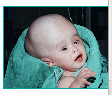
Hydrocephalus is the buildup of fluid in the cavities (ventricles) deep within the brain. The excess fluid increases the size of the ventricles and puts pressure on the brain. Cerebrospinal fluid normally flows through the ventricles and bathes the brain and spinal column. But the pressure of too much cerebrospinal fluid associated with hydrocephalus can damage brain tissues and cause a range of impairments in brain function. Hydrocephalus can happen at any age, but it occurs more frequently among infants and adults 60 and over. Surgical treatment for hydrocephalus can restore and maintain normal cerebrospinal fluid levels in the brain. Many different therapies are often required to manage symptoms or functional impairments resulting from hydrocephalus.